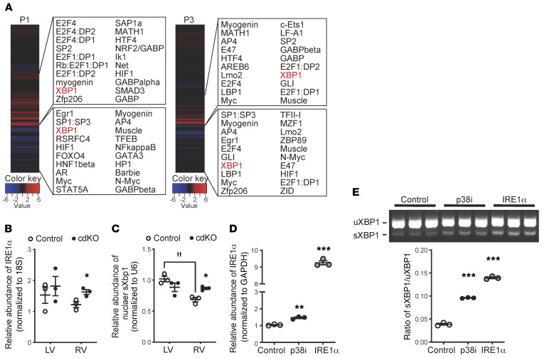
Figure 8. IRE1α/XBP1 and p38 MAPK activity in cardiomyocytes.
(A) Whole-genome rVISTA analysis of the differentially expressed genes in the P1 and P3 p38-cdKO RV vs. the control RV. The top 20 candidate transcription factors are listed for the upregulated genes (upper panels) and the downregulated genes (bottom panels). (B) Chamber-specific expression of IRE1α mRNA in the P3 control and p38-cdKO ventricles (n = 3; mean ± SEM). *P < 0.05 (vs. control). (C) Chamber-specific expression of nuclear sXbp1 mRNA at P3 in the control and the p38-cdKO ventricles (n = 3; mean ± SEM). *P < 0.05 (vs. control); ††P < 0.01 (LV vs. RV). (D) IRE1α expression in the rat neonatal ventricular myocytes (NRVMs) treated with p38 inhibitor (p38i, SB202190) or Adv-IRE1α (n = 3; mean ± SEM). (E) The unspliced (uXbp1) and spliced (sXbp1) Xbp1 mRNA levels in the NRVMs treated with p38i (SB202190) or Adv-IRE1α (n = 3; mean ± SEM). ***P < 0.001, **P < 0.01 (vs. control).

