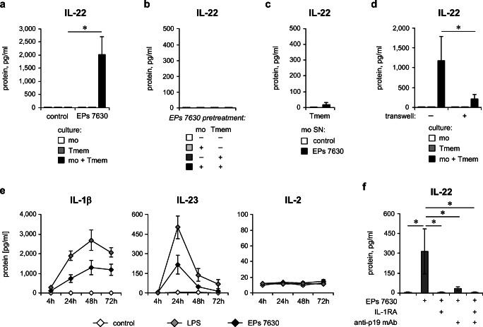
Fig. 5.
Monocytes play a key role in EPs® 7630–induced IL-22 production by T cells. a CD4+ memory T cells and autologous monocytes were co-cultured or cultured alone for 72 h in the absence (control) or presence of 10 μg/ml EPs® 7630 (EPs® 7630). b CD4+ memory T cells and autologous monocytes were pretreated in separate cultures with EPs® 7630 (10 μg/ml) or medium with solvent for 24 h. Afterwards, CD4+ memory T cells and monocytes were washed and co-cultured as indicated for 72 h without further EPs® 7630 stimulation. c CD4+ memory T cells were cultured for 72 h in the presence of supernatant (SN) obtained from cultures of EPs® 7630–stimulated (10 μg/ml) monocytes. d CD4+ memory T cells and autologous monocytes were co-cultured with (no transwell) or without enabled cell-cell contact (transwell) or were cultured separately for 72 h in the presence of 10 μg/ml EPs® 7630. a–d Human CD4+ memory T cells and autologous monocytes were each isolated by magnetic labeling–based cell sorting. Quantification of IL-22 in culture supernatants was carried out by ELISA. e Human PBMCs were stimulated or not (solvent control) in a kinetic approach with 10 μg/ml EPs® 7630 or 100 ng/ml LPS or were left without stimulation up to 72 h. Quantification of IL-1β, IL-23, and IL-2 levels in culture supernatant was performed by ELISA. f Human PBMCs were stimulated or not (solvent control) with 3 μg/ml EPs® 7630, in the presence of 1.5 μg/ml IL-1RA, 3 μg/ml anti-IL-23p19 antibody, or a combination thereof for 72 h. Quantification of IL-22 in culture supernatants was carried out by ELISA. Data from 6 (a), 2 (b), 4 (c), 5 (d) 3–4 (e), and 5 (f) independent experiments are given as mean ± SEM. Significant differences between treatment groups are indicated (*p < 0.05, Wilcoxon matched-pairs signed-rank test). Tmem: CD4+ memory T cells; mo: monocytes

