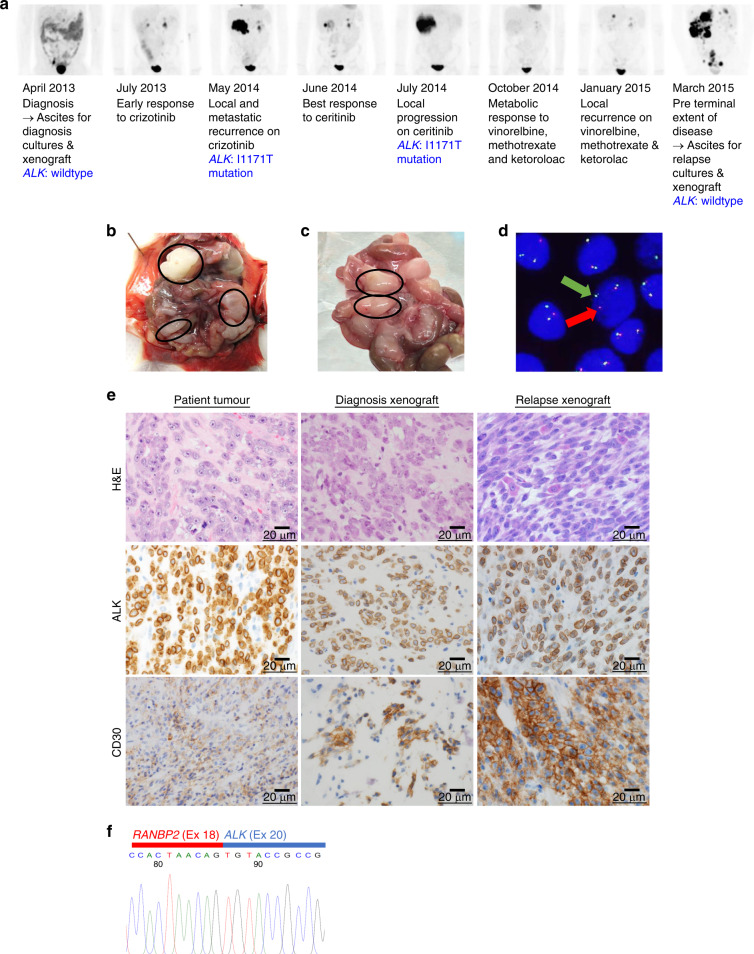
Fig. 1. eIMS xenografts recapitulate the features of the patient’s disease.
a The patient’s clinical response to successive treatments measured by FDG–PET scan and timepoints where patient samples were collected. The identification of the ALK-resistance mutation has been previously reported;9 xenografts were established by intraperitoneal injection of eIMS cells from cultured ascites harvested at b diagnosis and c relapse. Multinodular disease developed in the abdominal cavity of each inoculated mouse. Tumour nodules are circled. d Cell suspensions and short-term cultures of xenograft cells were analysed by FISH using a break-apart ALK locus probe. ALK gene rearrangement is indicated by the split signal (indicated by red and green arrows). A representative image of diagnosis xenograft cells shown. e Xenograft tumours retain histopathologic features of the patient tumour, including perinuclear ALK localisation and CD30 expression. Images ×600 magnification. f Sanger sequencing of xenograft RNA confirming an in-frame RANBP2-ALK fusion between exon 18 (Ex 18, red bar) of RANBP2 and exon 20 (Ex 20, blue bar).

