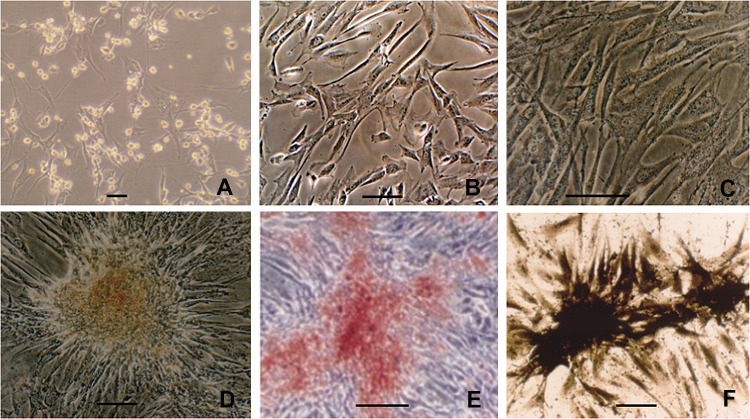
Fig. 2.
Seeding of cells. A MSCs of primary passage; B Uniform morphology of the 3rd passage MSCs; C confluent MSCs induced with osteogenic media showed intracellular refractive granules; D after 3-week induction, MSCs clused and formed calcified nodules; E calcified nodules formation from osteogenically-induced MSCs confirmed by alizarin red staining; F ALP expression of osteogenically differentiated MSCs visualized by the modified Gomori staining. Scale bar = 100 μm

