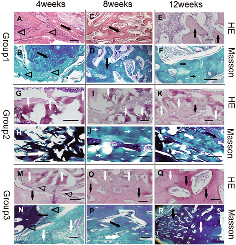
Fig. 6.
Histological examination under light microscope with HE (row 1, 3 and 5) and Masson’s trichrome (row 2, 4, and 6) staining. In Group 1 (row 1, 2), TEP formed island-like calluses at 4 weeks (A, B). New bone tissue increased with irregular vessels or immature marrow cavities, while TEP disappeared (possibly degraded) at 8 weeks (C, D). At 12 weeks, all newly-formed bone developed into mature cancellous bone (E, F). In Group 2 (rows 3, 4), DPB was mainly surrounded by scar tissue and infiltrated lymphocytes at 4 weeks (G, H), and accompanied by little new osseous tissue formation at 8 (I, J) or 12 weeks (K, L). In Group 3 (rows 5, 6), there was a small amount of new bone formation between TEP and DPB, which were degraded accompanied by scar tissue at 4 weeks (M, N). New osseous tissue formed woven bone at 8 weeks (O, P), while it was inclined to form mature lamellar bone with osteoblasts embedded into mineral matrix at 12 weeks (Q, R). Triangles represent TEP, black arrows represent newly-formed bone tissue, white arrows represent remnants of DPB. Scale bar = 1 mm

