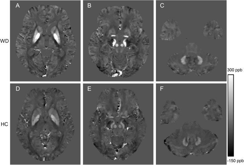
FIGURE 2.
Comparison of QSM images from a WD patient (36 years old, male, A–C) and a healthy control (38 years old, male, D–F). The QSM images (A,D) include head of the caudate nucleus, globus pallidus, putamen and thalamus. The QSM images (B,E) include the red nucleus and substantia nigra. The QSM images (C,F) include the dentate nucleus. Significantly increased susceptibility was observed in the deep gray matter nuclei of the WD patient.

