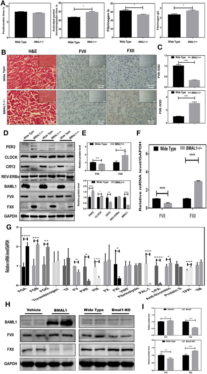
Figure 3.

Disrupted coagulation factors VII and XII in Bmal1−/− mice. (A) Routine coagulation array for whole blood in Bmal1−/− vs. WT mice (3/group). (B,C) H&E and IHC staining for coagulation factors VII and XII in slices of livers, and the relative values of IHC optical density. (D–G) Western blot analysis and qRT-PCR analysis of circadian clock genes and coagulation factors in mouse liver. (H,I) Western blot analysis of coagulation factors VII and XII in Bmal1-overexpression and Bmal1-knockout HepG2 cells. The experiments were performed three times independently. *P < 0.05; **P < 0.01; ***P < 0.005; ****P < 0.001.
