Figure 7.
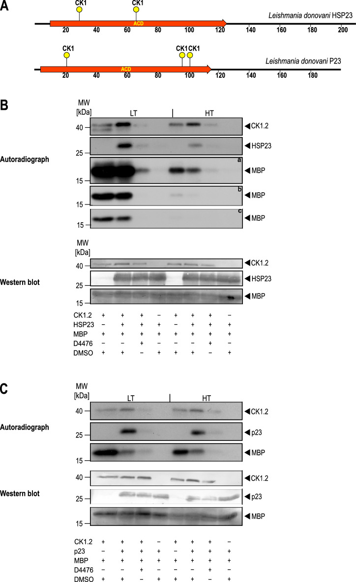
In vitro kinase assays using [γ-32P]-ATP. (A) Schematic representation of the putative CK1 phosphorylation sites present in HSP23 and P23. (B, C) 0.25 µg recombinant Casein kinase 1.2 was incubated with 3 µg of myelin basic protein (MBP), 1.57 µg of recombinant HSP23 (B) or 1.3 µg P23 (C) in the presence of either 10 µM D4476 inhibitor or equivalent concentrations of DMSO. The reactions were incubated at LT (25 °C) or HT (37 °C) for 30 min and stopped by addition of loading buffer. A reaction without the kinase was included to test for possible autophosphorylation of substrates. Proteins were separated on a 12.5% SDS-PAGE gel followed by Western blot. The incorporation of [γ-32P]-ATP was monitored by autoradiography (upper panels). a = 1 week exposure; b = 24 h exposure, c = 12 h exposure of X-Ray films. The blot was probed with an anti-CK1.2 (SY3535), HSP23 and P23 antibody, or stained with Coomassie blue to evaluate loading (lower panels). The figure shows representative Western blots from a series (n = 4) of experiments.

