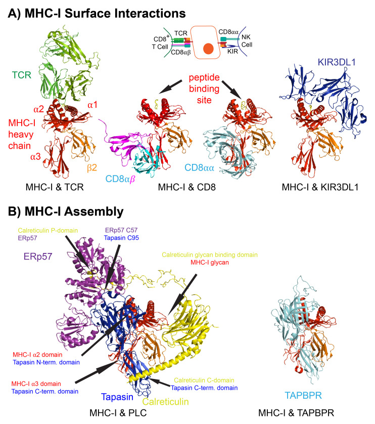
Figure 1. Major histocompatibility class I (MHC-I) surface interactions and assembly.
( A) Crystal structure and cartoon representation of MHC-I (red: heavy chain, orange: β2m, yellow: peptide)/TCR (green) (PDB 5C07 3) on CD8 + T cells, MHC-I/CD8 co-receptor (cyan and magenta) (PDB 3DMM 4 and PDB 3QZW 5), or MHC-I/KIR3DL1 (blue) on natural killer (NK) cells (PDB 5B38 6). ( B) Cryo-EM structure of MHC-I in the PLC (yellow: calreticulin, blue: tapasin, purple: ERp57) (PDB 6ENY 7 adapted from data freely accessible at: https://www.rcsb.org/structure/6ENY) or with TAPBPR (cyan) (PDB 5WER 8) in the peptide-deficient form. Arrows highlight interactions between tapasin and MHC-I, calreticulin and the MHC-I glycan, calreticulin and tapasin, calreticulin and ERp57, and tapasin and Erp57. β2m, beta2-microglobulin; KIR, killer cell immunoglobulin-like receptor; PLC, peptide loading complex; TAPBPR, transporter associated with antigen presentation-binding protein related; TCR, T-cell receptor.

