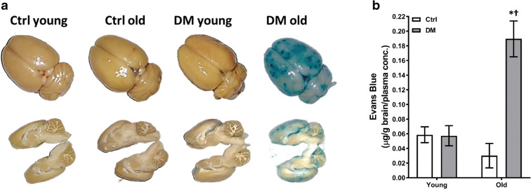
Fig. 3.
Blood-brain barrier (BBB) permeability. Comparison of BBB permeability in young and old non-diabetic control (Ctrl) and DM rats. a Representative images of the extravasation of Evans blue after mean blood pressure (MAP) was elevated to 180 mmHg for 1 h. b Tissue concentration of Evans blue in the brains. Mean values ± SEM are presented. N = 4–6 rats per group. The asterisk indicates P < 0.05 from the corresponding values in age-matched SD rats. The dagger indicates P < 0.05 from the corresponding values in young rats within a strain

