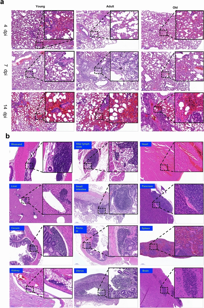
Figure 2.
Histopathological examination in SARS-CoV-2 infected tree shrews. On 4, 7 and 14/16 dpi, tree shrews were euthanized and dissected. Tissues were collected from each animal for H&E staining and histopathological analysis. The histological sites with typical and representative lesions were zoomed in, which was described in text. (a) Histological lesions in the pulmonary tissues. The representative images were shown here from young, adult and old tree shrews dissected at the different stages of SARS-CoV-2 infection. (b) Histopathological changes in the other twelve tissues. Each image here represents one tissue of an animal, including weasand, Hilar, heart, liver, small intestine, pancreas, cecum, rectum, spleen, kidney, uterus, brain. Histopathological score of tissues in all tree shrews was summarized in Table 3.

