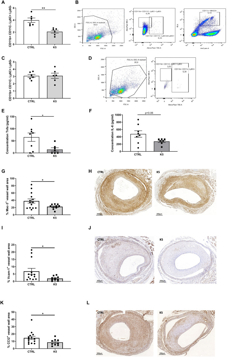
Figure 5.
K5 reduces circulating monocytes and macrophage infiltration in advanced vein graft atherosclerotic lesions in ApoE3*Leiden mice. (A) FACS quantification of circulating monocytes in the blood of control and K5 treated mice and (B) gating strategy (n = 6 mice per group). (C) FACS quantification of monocytes in the spleen of control and K5 treated mice and (D) gating strategy (n = 6 mice per group). Quantification of the concentration of Tnf (E) and IL-6 (F) in samples from whole blood assay incubated with LPS (n = 8 mice per group). (G) Quantification of Mac3 positive area in the control and K5 groups and (H) respective example of the staining on the right (n = 14 in CTRL group and n = 10 in K5 group). (I) Quantification of Vcam-1 and (K) Ccl-2 positive area in the control and K5 groups (n = 14 in CTRL group and n = 10 in K5 group). (J) Representative pictures for Vcam-1 IHC staining and (L) Ccl2 IHC staining in mice from ctrl and K5 groups (n = 14 in CTRL group and n = 10 in K5 group). Data are presented as mean ± SEM. *p < 0.05, **p < 0.01, ***p < 0.001. ****p < 0.0001; by 2-sided Student t test.

