FIGURE 1.

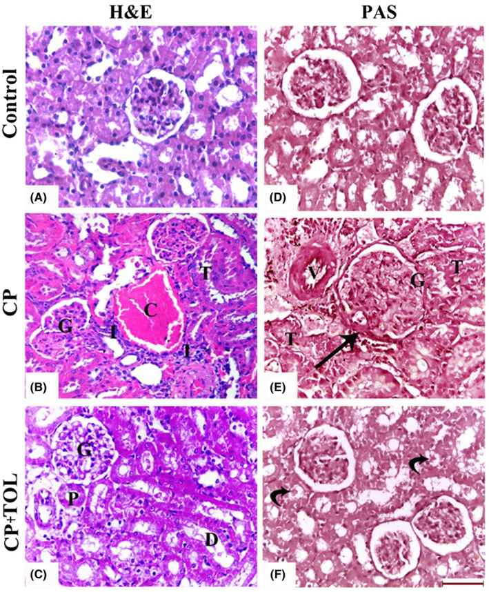
H&E‐stained sections of (A): Control group, (B) CP‐treated group showing marked congestion (C) of the blood vessels and inflammatory cellular infiltration (I). Note: distortion of the tubules (T) and glomeruli (G), (C) CP + TOL‐treated group showing improvement in the structural pattern of the glomeruli (G), proximal (P), and distal (D) convoluted tubules, Periodic Acid‐Schiff (PAS)‐stained sections of (D): Control group, (E) CP‐treated group showing thickening of the Bowman's capsule (arrow) and wall of blood vessels (V). The renal tubules (T) show disruption of their basal laminae and appear obliterated with casts. (F), CP + TOL‐treated group showing normal glomerular PAS reaction. The tubules exhibit few casts (curved arrows; (Scale bar 50 μm)
