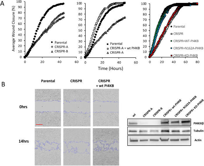
FIGURE 3:
Loss of PI4KIIIβ impairs migration. (A) (Left panel) The ability to close an in vitro wound is shown in two independent lines of PI4KIIIβ-deleted cells compared with parental cells. (Center panel) Attenuated wound healing is rescued in the CRISPR line by re-expression of WT PI4KIIIβ (wtPI4KB). (Right panel) Attenuated wound healing is rescued in the CRISPR line by re-expression of wild-type PI4KIIIβ (WT-PI4KB) and KD (KD-PI4KB) but not a Rab11-binding mutant (N162A-PI4KB). Experiments are representative of triplicates. (B) (Left panels) Inset shows a representative image from the wound closure experiment in the center panel of A. Scale bar in red is 300 μm. (Right panel). Protein expression of PI4KIIIβ and tubulin and actin control in the cell lines.

