FIGURE 5:
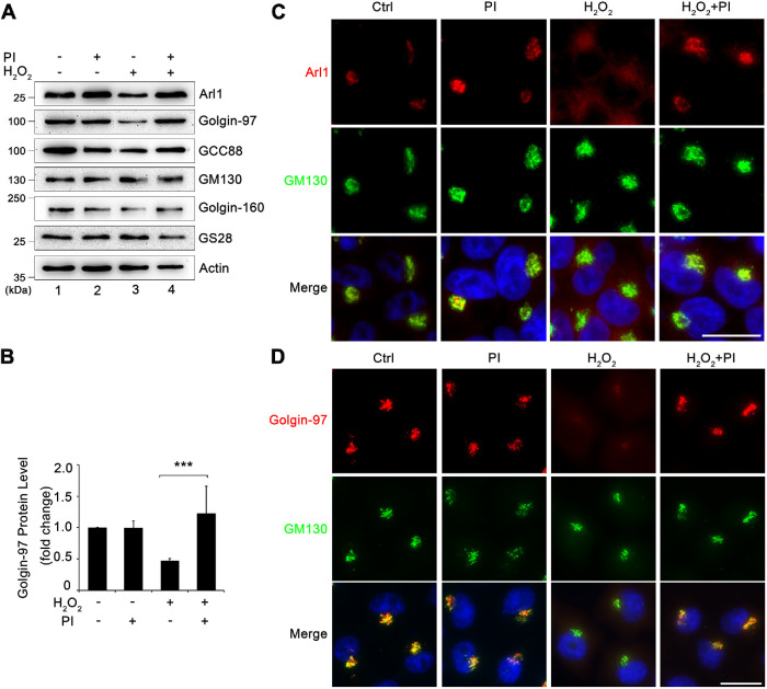
H2O2 treatment causes Arl1 and Golgin-97 degradation by cytoplasmic proteases. (A) HeLa cells cotreated with or without a protease inhibitor cocktail and 1 mM H2O2 for 10 min were analyzed by Western blotting. (B) Quantitation of Golgin-97 Western blot in A with densitometric analysis, with the control normalized to 1. Results are shown as mean ± SEM from three independent experiments. Statistical analyses were performed using two-tailed Student’s t tests (***, p ≤ 0.001). (C) Cells treated as in A were stained for Arl1 (red), GM130 (green), and Hoechst (blue). (D) Cells treated as in A were stained for Golgin-97 (red), GM130 (green), and DNA (blue). Scale bars = 20 µm.

