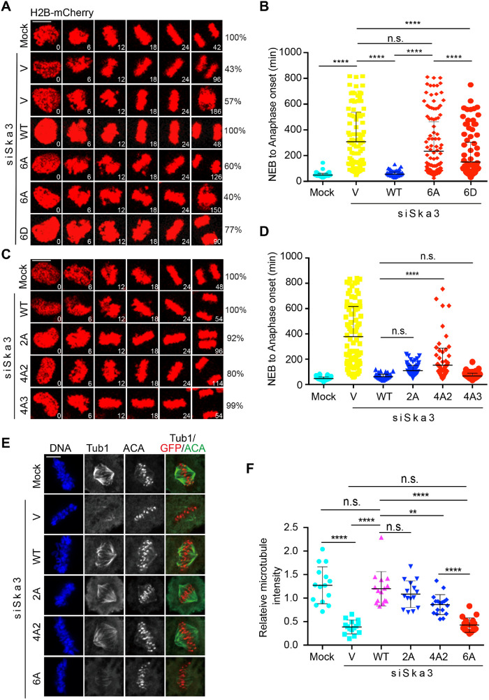
FIGURE 4:
Cdk1 phosphorylation in Ska3 is essential for chromosome segregation. (A, C) HeLa Tet-On cells stably expressing H2B-mCherry were cotransfected with siSka3 and vectors (V) or plasmids containing GFP-Ska3 WT, 2A (C), 4A2 (C), 4A3 (C), 6A (A), or 6D (A). Time-lapse microscopic analysis was performed. Scale bars, 5 μm. (B, D) Quantification of the duration from NEB to anaphase onset in A and C. At least 100 mitotic cells in A and 80 mitotic cells in C were analyzed for each condition. Average and SD were shown in lines. *P < 0.05, **P < 0.01; ****P < 0.0001; n.s. denotes no significance. Scale bars, 5 μm. (E) Cdk1 phosphorylation in Ska3 is required for stability of kinetochore–microtubule attachments. siSka3-treated HeLa Tet-On cells transfected with vectors or plasmids containing GFP-Ska3 WT, 2A, 4A2, or 6A were treated with MG132 for 1 h after 9 h release from thymidine, incubated on ice for 5 min, and then subject to staining with DAPI and the indicated antibodies. Representative images are shown here. (F) Quantification of microtubule intensity in E normalized to that of DNA. At least 15 mitotic cells (10 microtubules per cell) were analyzed for each condition. Average and SD are shown in lines. **P < 0.01; ****P < 0.0001; n.s. denotes no significance.

