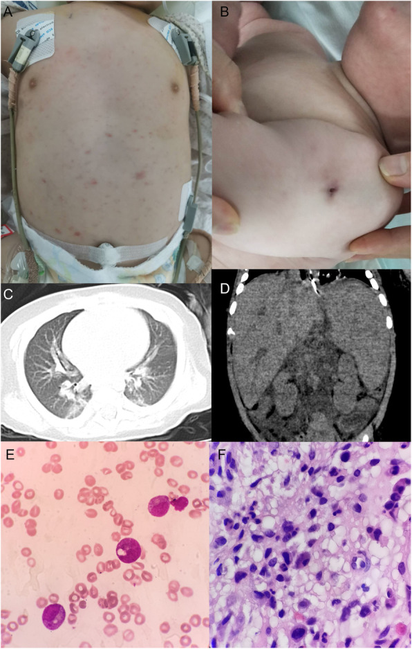
Fig. 1.

a Papule on the trunk. b Unhealed BCG scar on the left arm. c Chest CT scan revealed infectious lesions scattered in lungs. d Abdomen CT scan showed enlarged liver and spleen. e No hemophagocytosis was observed by bone marrow aspiration via hematoxylin and eosin (H&E) staining, high power view (× 400), lack of lymphocytes, but toxic granulation and vacuolus in neutrophils, which are signs of severe infection. f Numerous inflammatory cells, stained via H&E staining, high power view (× 400), were shown by bone marrow biopsy, but without evidence of malignancy
