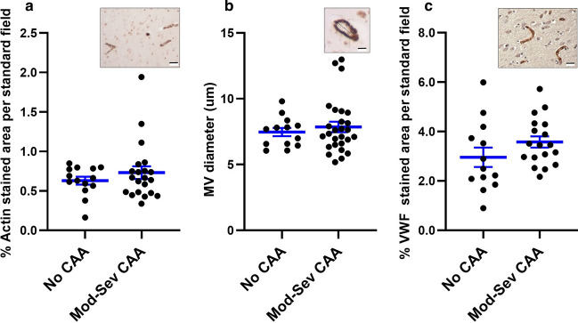
Fig. 2.
Density and diameter of MV are not significantly different in samples of occipital cortex with no and moderate-severe CAA pathology. Shown are scatter plots with mean and SEM. Density (a, c) and diameter (b) of MV were measured on samples labeled by IHC for alpha-smooth muscle actin (a, b with insets of representative images) and vWF (c with inset of representative image). These assays show no significant differences in MV density and diameter among the three groups. Inset scale bars are 20 µm (a, c) and 5 µm (b). Sample sizes (n) are indicated in “Results”

