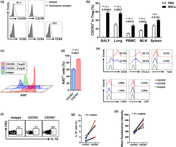
Figure 4.

MSC‐induced Tregs highly express chemokine receptor CXCR3 and exhibit enhanced anti‐inflammatory functions. Mice were injected with MSCs or PBS. Three days later, mice were sacrificed. (a) Expression of chemokine receptors by lung Tregs was evaluated, and representative FACS graphs were shown. (b) Frequencies of CXCR3+ Tregs in lung and peripheral lymphoid tissues were determined. n = 3 per group. (c, d) Proliferative ability of CXCR3− and CXCR3+ Tregs was compared. Expression of ki67 was measured. n = 5 per group. (e) Expression of ICOS, CTLA‐4, T‐bet, LAG3 and LAP was examined. Production of IL‐10 by CXCR3− and CXCR3+ Tregs from MSC‐treated mice was determined by intracellular staining. (f) Representative FACS data. (g) Percentages of IL‐10‐producing Tregs. (h) Mean fluorescence intensity of IL‐10. n = 5 per group. All the experiments were repeated three times. Representative data are shown.
