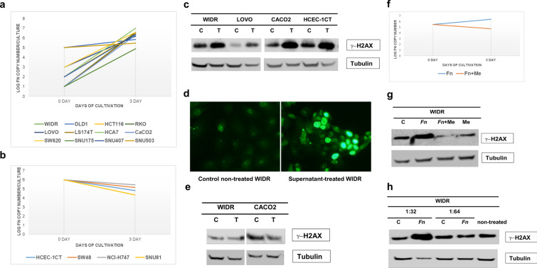
Fig. 3.
Aerobic growth of Fn and induction of γ-H2AX by Fn. (a) Twelve colon cancer cell lines (WIDR, DLD1, HCT116, RKO, LOVO, LS174T, HCA7, CaCO2, SW620, SNU175, SNU407, and SNU503) were infected with Fn strain EAVG_002. During a 3-day incubation, an increase in Fn copy number was observed in co-cultures with these cell lines. (b) No increase in Fn copy number was observed in co-cultures with 4 different colon cancer cell lines (HCEC-1CT, SW48, NCI-H747 and SNU81). X-axis: days of cultivation; Y-axis: log of Fn copy number per culture. (c) WIDR cells were infected with Fn EAVG_002 strain at a multiplicity of infection of 1 under 5%CO2/21%O2 for 1 week. Supernatants were collected, centrifuged, and filtered through a 0.2 μm porous membrane. 2 × 10⁵ cells (WIDR, LOVO, CaCO2 and HCEC-1CT) were exposed to the supernatants for 9 hr. Cell lysates were analyzed for induction of γ-H2AX by Western blotting using anti-γ-H2AX mouse monoclonal antibodies. C: control non-treated; T: supernatant treated. Treated cells expressed more γ-H2AX than control non-treated cells. (d) Immunofluorescent staining of Fn supernatant-treated WIDR cells with anti-γ-H2AX mouse monoclonal antibodies. Brighter nuclear γ-H2AX signal is evident in treated cells. (e) Supernatant from HCEC-1CT cell culture infected with Fn EAVG_002 strain at an MOI of 1 under 5%CO2/21%O2 conditions failed to induce γ-H2AX in supernatant-treated WIDR and CaCO2 cells. C: control non-treated; T: supernatant treated. There was no difference in the amount of γ-H2AX between control non-treated and supernatant treated cells. (f) Treatment of co-culture between WIDR and Fn with metronidazole inhibited Fn aerobic growth (red line). Blue line represents growth of Fn without metronidazole. (g) Metronidazole abolished γ-H2AX induction by Fn. Supernatants were collected from 4 cultures (C: control non-treated cells; Fn: Fn infected cells; Fn + Me: Fn infected cells treated with metronidazole; Me: non-infected cells treated with metronidazole alone) and tested for induction of γ-H2AX in WIDR cells. (h) Bacterial medium in which Fn grew anaerobically was tested for γ-H2AX induction. The Fn grown medium and control fresh bacterial medium was diluted by Dulbecco's modified Eagle medium with 10% fetal bovine serum at 1:32 and 1:64 ratio and then exposed to WIDR cells. C: WIDR cells were treated with a diluted fresh bacterial medium; Fn: WIDR cells were treated with diluted Fn grown medium. Induction of γ-H2AX was detected at 1:32 but not at 1:64 dilutions of Fn grown medium

