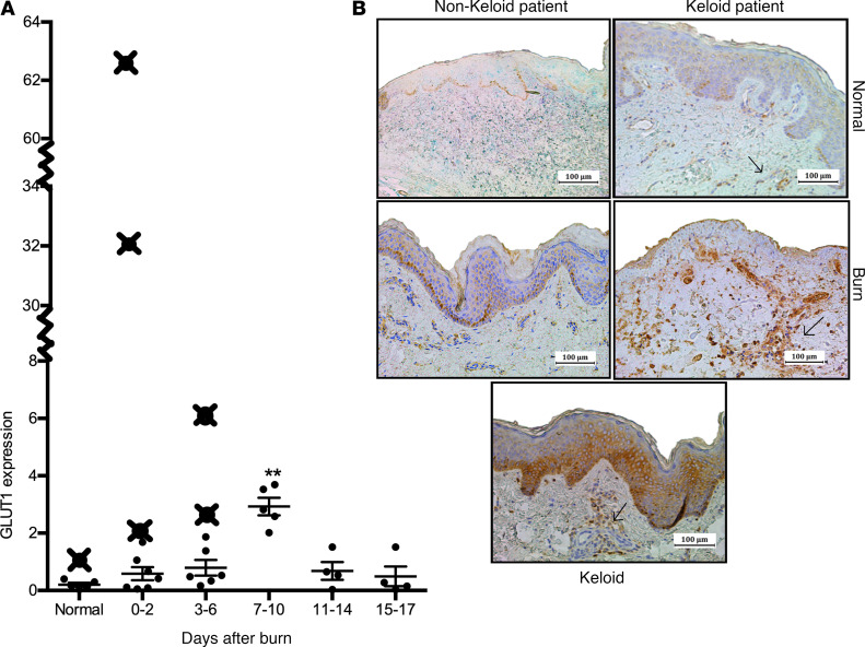
Figure 2. Burn patients who develop keloids have prior indications of altered glucose metabolism.
(A) GLUT1 gene expression in burn and normal skin samples obtained from burn patients before the development of keloids indicates higher expression compared with typical values seen in nonkeloid patients (≥ 2 SDs above mean). Similarly, keloids exhibited increased Glut1 staining. (B) Immunohistochemical staining for Glut1 in skin from keloid patients indicates more Glut1+ cells in basal epidermal and dermal layers compared with nonkeloid counterparts (normal skin, top panel; burn skin 7–10 days after burn, bottom panel). Individual data points (X) denote gene expression values for individual patients. Values are presented as mean ± SEM. Experiments were conducted twice. One-way ANOVA; **P < 0.01 burn versus normal.

