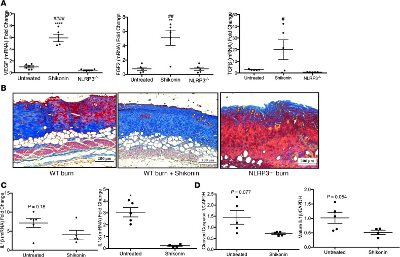
Figure 5. Shikonin treatment is not detrimental to wound healing in vivo.
(A) Gene expression of VEGF, FGF2, and TGFβ at 7 days after burn (n = 5–6). (B) Trichrome staining of excised burn skin from untreated WT burn (left), treated WT burn (center), and untreated NLRP3–/– burn (right) mice indicates increased dermal collagen deposition–treated mice. (C) Murine skin gene expression for IL1β (n = 5–6) and IL18 (n = 4–6) at 7 days after burn. (D) Protein expression for cleaved caspase-1 and mature IL-1β in untreated and shikonin-treated murine skin (n = 4–5). Values are expressed as average fold change relative to normal skin, presented as mean ± SEM. Experiments were conducted twice. One-way ANOVA with Tukey’s post hoc or Students t test; *P < 0.05 and **P < 0.01; ***P < 0.001; ****P < 0.0001 shikonin versus untreated; #P < 0.05, ##P < 0.01, ###P < 0.001, and ####P < 0.0001 shikonin versus NLRP3–/–.

