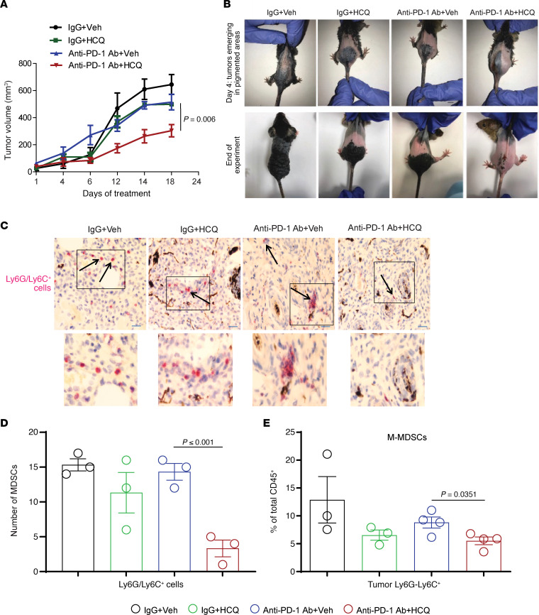
Figure 4. HCQ and anti–PD-1 Ab combination impairs tumor growth and reduces the MDSCs’ infiltration in a BRafCA PtenloxP Tyr::CreERT2 melanoma model.
(A) Topical 4-HT was applied on the back to elicit spontaneous melanoma growth (n = 4 per treatment), and once tumors were palpable treatment as in Figure 1 was started. (B) Representative images of mice. (C) Representative images of IHC staining of tumor against Ly6C/Ly6G (MDSC marker) at original magnification ×10 (inset of each image is ×2.5 magnified). (D) Number of Ly6C/Ly6G+ cells per high-powered field. (E) Immunophenotyping of M-MDSCs in tumor. A P value is presented for the test of the hypothesis that the addition of HCQ to anti–PD-1 Ab is significantly different compared with anti–PD-1 Ab + Veh. All t tests were 2 tailed and 2 sample.

