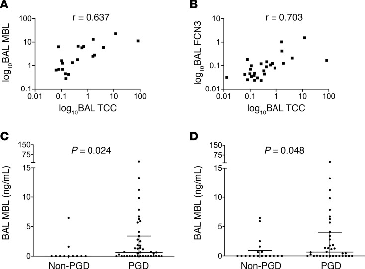
Figure 5. Local markers of lectin pathway activation distinguish subjects with PGD.
Levels of mannose-binding lectin (MBL) in the bronchoalveolar lavage (BAL) highly correlated with markers of complement activation in the BAL (soluble terminal complement complex [TCC]) in the WUSM cohort (A, n = 40). Using a different assay than in Figure 4 (n = 73), BAL MBL levels were higher in subjects who developed PGD compared with the levels in subjects without PGD (C), and this held true in those who developed PGD at or after 24 hours (D). The levels of ficolin-3 (FCN3; B) also highly correlated with BAL TCC (n = 40). r represents Spearman’s rho coefficient. The axes were expressed in a logarithmic scale for purposes of graphical representation. Rank sum tests of comparison (Mann-Whitney U test).

