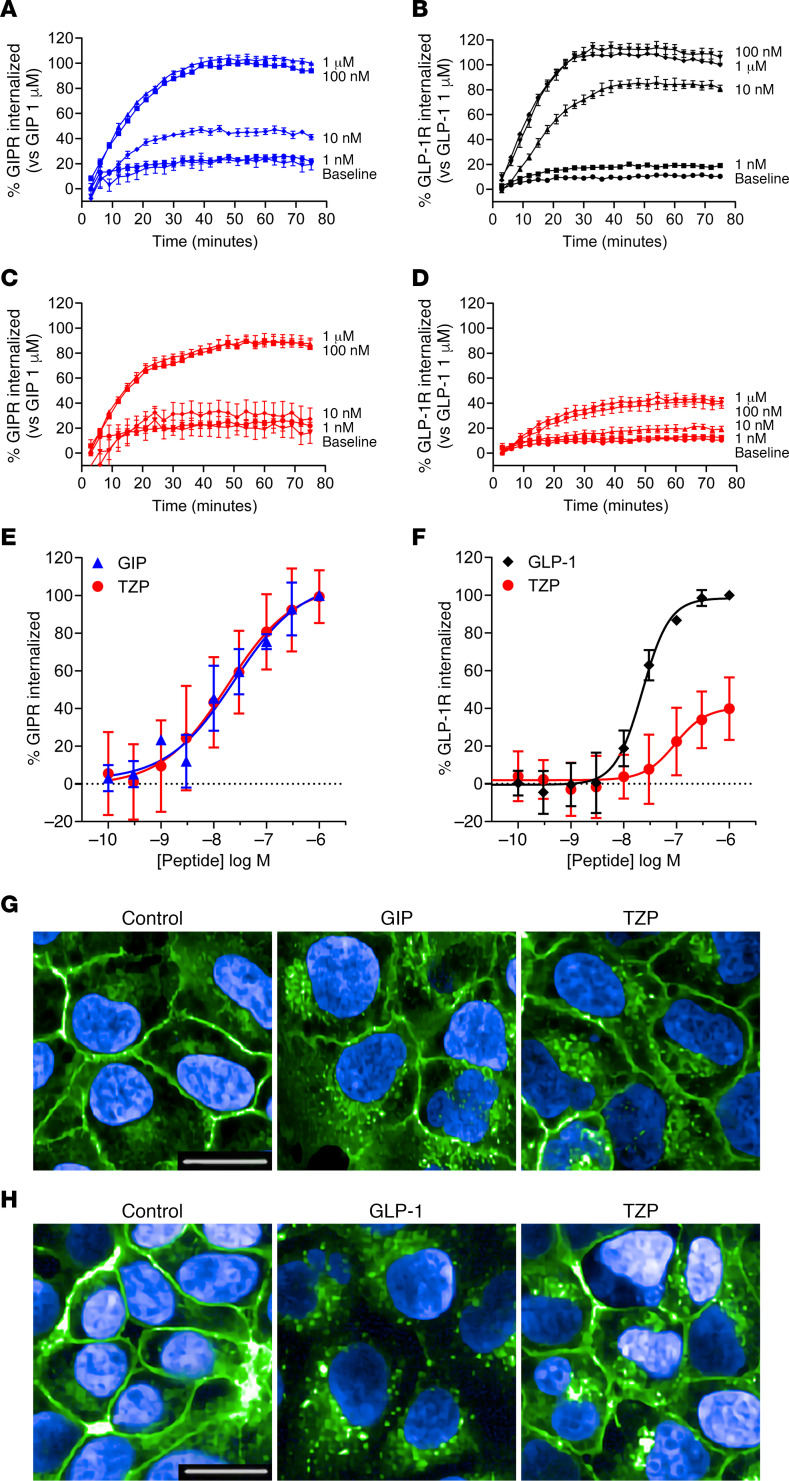
Figure 2. Tirzepatide (TZP) differentially induces internalization of the GIP and GLP-1 receptors.
(A–D) The time course of internalization of GIPR (A and C) and GLP-1R (B and D) was assessed using changes in cell surface presentation of SNAP-tagged receptors in HEK293A cells. Receptor internalization induced by increasing concentrations of GIP(1-42) (A), GLP-1(7-36) (B), or tirzepatide (C, GIPR; D, GLP-1R) over time relative to the maximum signal for either GIP(1-42) (1 μM) or GLP-1(7-36) (1 μM) is shown. (E–H) Studies using receptors containing an N-terminal HA-epitope tag and a C-terminal EGFP fusion are presented. (E) Changes in surface GIPR 60 minutes after treatment with ligand were measured by anti-HA immunofluorescence. Data are normalized to 1 μM GIP(1-42) values. For GIPR, tirzepatide induced internalization with an EC50 (SEM, n) of 18.1 nM (5.7, 4), while GIP(1-42) displayed a potency of 18.2 nM (9.7, 4). (G) Representative confocal images of HA-GIPR-EGFP cells detecting EGFP fluorescence following treatment with vehicle, 100 nM GIP(1-42), or 100 nM tirzepatide. (F) Changes in surface GLP-1R 30 minutes after treatment with ligand detected by anti-HA immunofluorescence. Data are normalized to 1 μM GLP-1(7-36) values. For GLP-1R, tirzepatide was partially efficacious at 43.6% (7.9, 3) with an EC50 of 101.9 nM (29.8, 3), while GLP-1(7-36) showed a potency of 22.2 nM (1.86, 3). (H) Representative confocal images of HA–GLP-1R–EGFP cells detecting EGFP fluorescence following treatment with vehicle, 100 nM GLP-1(7-36), or 100 nM tirzepatide. Scale bars: 20 μm.

