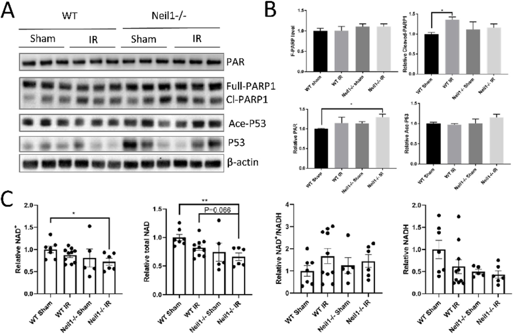
Fig. 6.
DNA damage is increased in Neil1−/− after 4 weeks of IR exposure. Representative immunoblots of the indicated proteins from the hippocampi of WT, and Neil1 −/− after 4 weeks of IR exposure (A). Quantification of immunoblots of the PAR, Ace-P53, Cleave-PARP1, Full-PARP1 proteins level (B). Relative proteins were normalized with β-actin. n = 3 mice per group. Data shown are mean ± SEM. *P < 0.05, **P < 0.01, ***P < 0.001. NAD+, total NAD, NADH, and NAD +/NADH ratio in cortex tissue of WT, Neil1−/− at 4 weeks after IR exposure (C). n=7 WT sham, 10 WT IR, 5 Neil1−/− sham, 6 Neil1−/− IR mice. Data shown are mean ± SEM. *P < 0.05, **P < 0.01, ***P < 0.001.

