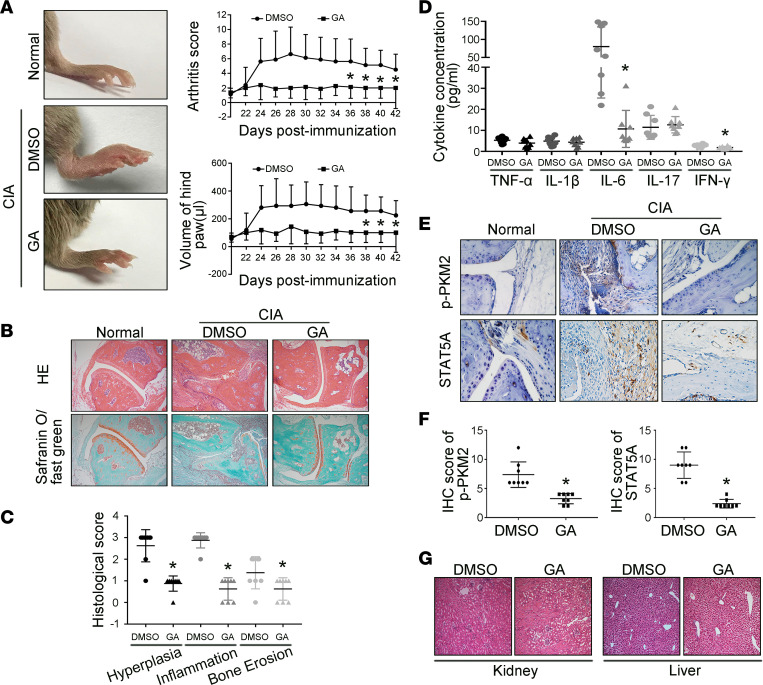
Figure 6. Effect of the SAE1/UBA2 inhibitor GA on the severity of arthritis in mice with CIA.
DBA/1J male mice were immunized with bovine type II collagen in complete Freund’s adjuvant and administered a booster injection 21 days later to induce CIA. On the 22nd day, the mice were injected i.p. with GA (30 mg/kg/d) or DMSO (as a model control) daily for 21 days. (A) Effect of GA on clinical scores and paw swelling (changes in volume) in CIA mice. The values in A are the mean ± SD in 8 mice treated with GA or 8 mice treated with DMSO. (B and C) Histological appearance of the joints of normal control and CIA mice treated with DMSO (n = 8) or GA (n = 8). H&E staining was used to evaluate synovial infiltration, cartilage erosion, and bone loss, whereas the lower panel shows safranin O/fast green staining demonstrating proteoglycan depletion (B). Original magnification, ×100. The scores for synovial inflammation, cartilage erosion, proteoglycan depletion, and bone loss are shown as the mean ± SD in C. (D) Effect of GA treatment on levels of cytokines in ankle tissues of CIA mice. The concentration of cytokines was measured with the Milliplex Map Mouse Cytokine Kit. (E and F) Expression of p-PKM2 and STAT5A, measured by immunohistochemical staining, in synovial tissue from CIA mice. Representative images (E) and quantification of the percentage of p-PKM2–positive and STAT5A-positive cells (F) of normal mice (n = 5), DMSO-treated mice (n = 8), and GA-treated (n = 8) mice (original magnification, ×400). (G) Effect of GA on the kidney and liver of mice with CIA. Photomicrographs show the histopathology of the kidney and liver in mice treated with GA or DMSO. Original magnification, ×100. *P < 0.05 versus DMSO, Student’s t test (for C, D, and F) or 2-way ANOVA (for A).

