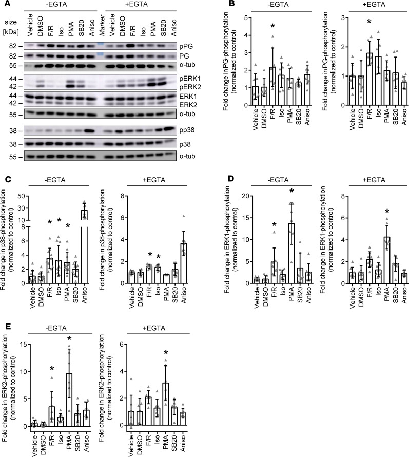
Figure 2. Phosphorylation of signaling proteins in basal and Ca2+-depleted conditions after treatments with mediators regulating cardiomyocyte cohesion.
(A) Representative Western blot of HL-1 cells treated with F/R, Iso, PMA, SB20, or Aniso under basal and Ca2+-depleted conditions. DMSO serves as control for SB20. (B–E) Quantification of changes in phosphorylation levels of (B) PG, (C) p38MAPK, (D) ERK1, and (E) ERK2, as compared with the respective controls upon treatment with mediators. *P ≤ 0.05, 1-way ANOVA with Bonferroni correction, n = 6–9. In C, Aniso was excluded from the statistical analysis, as the effect was so strong that all other effects were not significant anymore with 1-way ANOVA.

