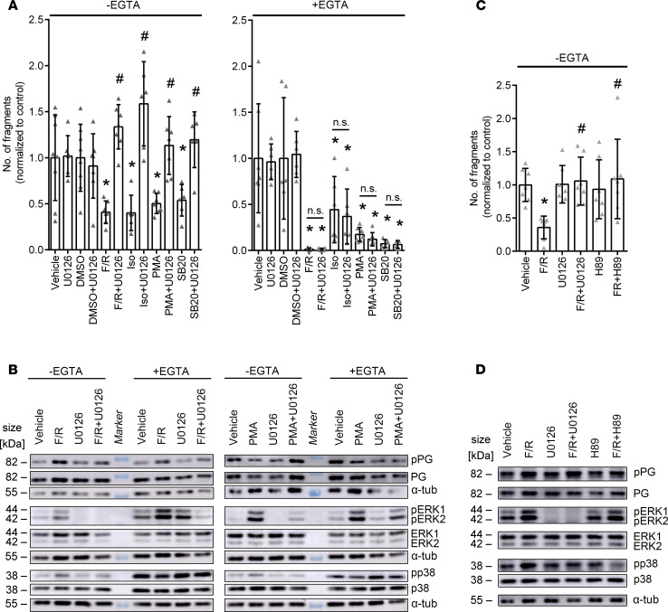
Figure 3. Inhibition of ERK1/2 alters cardiomyocyte cohesion in basal conditions and results in alteration of phosphorylation of PG, ERK1/2, and p38MAPK upon treatment with F/R and PMA.
(A) Dissociation assays in HL-1 cells treated with F/R, Iso, SB20, or PMA, with and without U0126 under basal and Ca2+-depleted conditions. DMSO serves as control for SB20. *P ≤ 0.05 as compared with the respective control, #P ≤ 0.05 as compared with the respective U0126-untreated condition, 1-way ANOVA with Holm-Šidák correction, n = 6. (B) Representative Western blot of HL-1 cells treated with F/R or PMA, with and without U0126 under basal and Ca2+-depleted conditions. n = 6. (C) Dissociation assays in HL-1 cells treated with F/R with and without U0126 or H89. *P ≤ 0.05 as compared with the vehicle control, #P ≤ 0.05 as compared with F/R, 1-way ANOVA with Holm-Šidák correction, n = 6. (D) Representative Western blot of HL-1 cells treated with F/R with and without U0126 or H89. n = 6.

