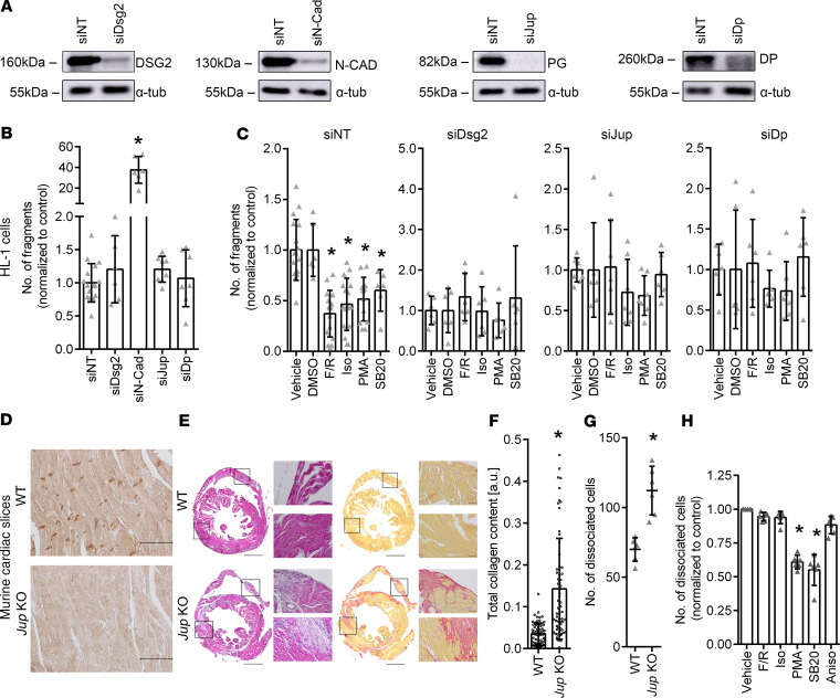
Figure 5. Effect of Dsg2, N-Cad, Jup, or Dp knockdown on F/R-, Iso-, PMA-, or SB20-mediated increase in cardiomyocyte cohesion.
(A) Representative Western blots confirming decreased levels of DSG2, N-CAD, PG, and DP after siRNA-mediated knockdown. (B) Dissociation assay under basal conditions following siNT, siDsg2, siN-Cad, siJup, and siDp treatments. *P ≤ 0.05, 1-way ANOVA with Holm-Šidák correction, n = 6. (C) Dissociation assays showing fold changes in fragments as compared with the respective controls in HL-1 cells following siNT, siDsg2, siJup, or siDp treatments and administration of F/R, Iso, PMA, or SB20. DMSO serves as control for SB20. *P ≤ 0.05, 1-way ANOVAs with Holm-Šidák correction, n = 6. (D) Immunohistochemistry of ventricular cardiac slices obtained from WT and PG-deficient mice (Jup-KO) stained for PG protein. Scale bar: 50 μm. n = 6. (E) H&E and Picrosirius red stainings of cardiac slices obtained from WT and Jup-KO mice. Scale bar: 1 mm; 50 µm (high-magnification views). (F) Total collagen content of cardiac slices obtained from WT and Jup-KO mice. *P ≤ 0.05, Student’s t test with Welch correction, n = 6. (G) Dissociation assays in murine cardiac slice cultures comparing the number of dissociated cells in WT and Jup-KO cardiac slices. *P ≤ 0.05, unpaired Student’s t test, n = 6. (H) Dissociation assays in murine cardiac slices obtained from Jup-KO mice following F/R, Iso, PMA, SB20, and Aniso treatments. *P ≤ 0.05, 1-way ANOVA with Holm-Šidák correction, n = 6.

