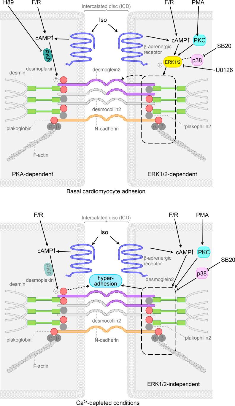
Figure 6. Schematic overview of signaling mechanisms involved in cohesion in HL-1 cardiomyocytes.
Adrenergic signaling (F/R and Iso), PKC activation (PMA), and p38MAPK inhibition (SB20) enhanced basal cardiomyocyte cohesion, referred to as positive adhesiotropy, in an ERK1/2-dependent manner. Positive adhesiotropy is possibly achieved through alterations in the interactions of the desmosomal proteins PG, PKP2, and DP (represented by dashed rectangle) that eventually lead to increased DSG2 translocation to the cell borders (represented by dashed arrow) and finally to positive adhesiotropy. On the other hand adrenergic signaling also acts via PKA-dependent PG phosphorylation at S665 and thereby increases basal cardiomyocyte cohesion. Under Ca2+-depleted conditions, adrenergic signaling, PKC activation, and p38MAPK inhibition lead to hyperadhesion independent of ERK1/2 (represented by dashed rectangle and straight arrow). Under the same conditions adrenergic signaling also induces PG phosphorylation, probably via PKA (shaded PKA), and thereby hyperadhesion (dashed arrow).

