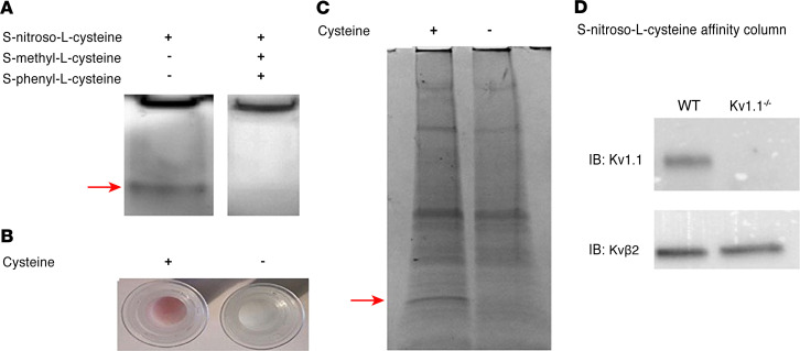
Figure 1. Identification of voltage-gated K+ channel proteins as binding partners for L-CSNO.
(A) Method 1. Murine cortex membrane proteins (50 μg/ lane; 2 sets of experiments using 1 C57BL/6 WT mouse) underwent native PAGE, then were incubated (30 minutes; dark; 27°C) with L-CSNO (50 μM) with or without 100 μM each L-CSMe and L-CSφ. Rinsed gels were developed with 40 μM DAF2 (30 minutes; dark; 27°C), and fluorescing bands (arrow) analyzed by LC-MS (compared with the control lane). Note that, because this is native PAGE, proteins were not separated before electrophoresis. (B) Method 2. Cysteine (100 mM) coupled to AminoLink Plus resin (4 hours, 27°C) or resin alone was incubated with EtONO (10 minutes; dark; 27°C). Pink color demonstrates S-nitrosothiol formation on the column (37). (C) Membrane proteins as in A (different animal) were loaded on columns (B) (30 min; dark; 27°C), washed, then eluted with Laemmli buffer followed by 0.1 M glycine, pH 3.5. Eluate underwent SDS-PAGE, and bands (including that shown by the arrow, 20 kDa) were analyzed by LC-MS in comparison with the control lane. Both methods identified several Kv channel proteins (see Supplemental Tables 2 and 3). Note that, because native PAGE was used (proteins were not separated before electrophoresis) in A, and a broad region of discordance was excised in C, multiple molecular weight proteins were evident by LC-MS. (D) Proteins as in A from WT and Kv1.1–/– mice (2 sets of experiments using WT and C57BL/6 background mice) were loaded on and eluted from columns as in B and C, and immunoblotted for Kv1.1 and Kvβ2.

