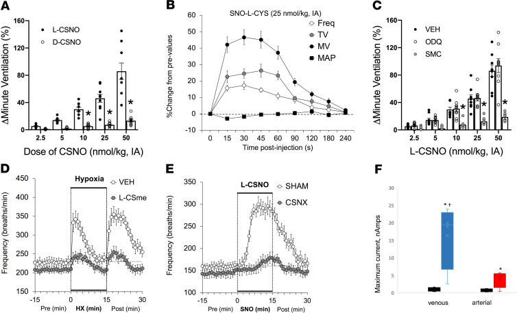
Figure 5. Stereoselective biological activity of the endogenous ligand, L-CSNO.
(A) Maximal increases in VE elicited by arterial injections of L-CSNO and D-CSNO in conscious Sprague-Dawley rats (n = 9). *P < 0.05, by ANOVA on ranks; †P < 0.05, D-CSNO versus L-CSNO. Mean ± SEM. IA, intra-arterial. (B) Changes in frequency of breathing (Freq), TV, VE [minute ventilation, MV]), and MAP elicited by a 25 nmol/kg dose of L-CSNO (n = 9 each; mean ± SEM at each time point). (C) Maximal increases in VE elicited by L-CSNO in Sprague-Dawley rats receiving infusions of vehicle (VEH; 0.1% DMSO in saline, 20 μL/min), ODQ (2 mg/kg bolus followed by 50 μg/kg/min, i.v.), or S-methyl-L-cysteine (SMC or L-CSMe) (10 μmol/kg/min, i.v.). n = 8–10 animals/experiment. *P < 0.05, SMC versus VEH or ODQ. Mean ± SEM. (D) Mice preinstrumented with arterial catheters at the level of the CB were treated with the L-CSNO congener L-CSMe (filled circles) or vehicle (open circles), then exposed sequentially to 10% oxygen or room air. L-SMC almost completely ablated the normal hypoxic response and recovery (n = 9 each; P < 0.001). At each time point, mean ± SEM. (E) The effect of L-CSNO is mediated by the CB. Mice underwent CSN (CSNO transection and carotid artery cannulation (as in Figure 5D). After a 3 week recovery, they were exposed to infusions of L-CSNO. Mice with intact CSN (SHAM, open circles) had a normal response to carotid L-CSNO infusion, while those with CSN transection (closed circles) did not (n = 12 each; mean ± SEM; P < 0.001 by ANOVA on ranks). (F) Detection of L-CSNO in blood samples. n = 3 each; median and CI. *P < 0.05, significant value; †P < 0.05, venous versus arterial, by ANOVA on ranks.

