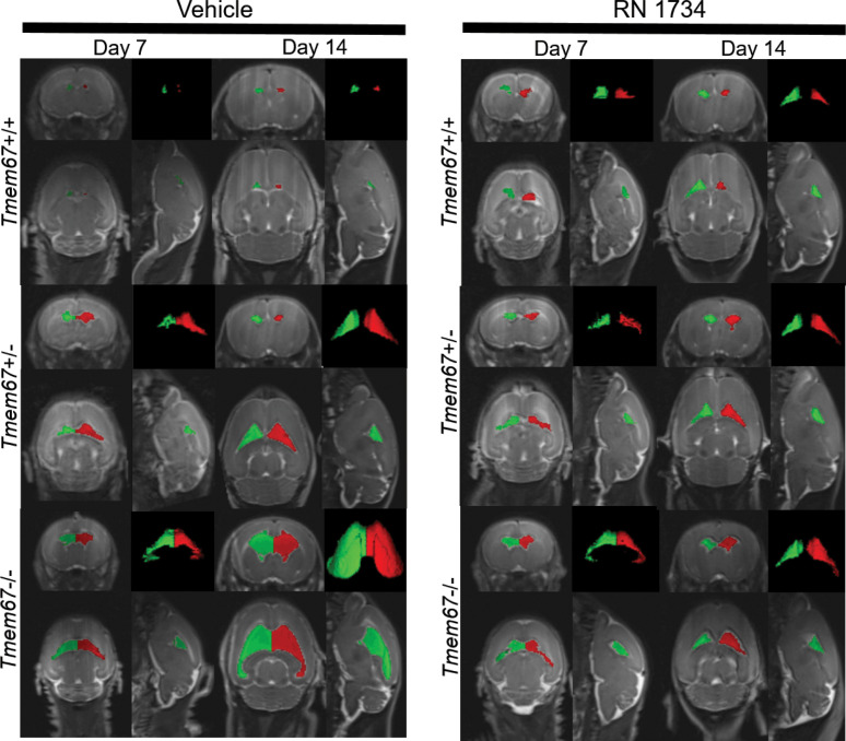
Figure 3. Representative MRI Scans Before and After Treatment with RN 1734.
MRI Images of P7 and P14 WT (Tmem67+/+), heterozygous (Tmem67+/–), and homozygous/hydrocephalic (Tmem67–/–) rats demonstrating the size of the lateral ventricles before and after treatment with vehicle or RN 1734. The images are shown as coronal, sagittal, and horizontal plane images, and a 3D rendering of the lateral ventricles. Red and green are pseudocolors of the right and left lateral ventricles to provide additional definition of the fluid compartments.

