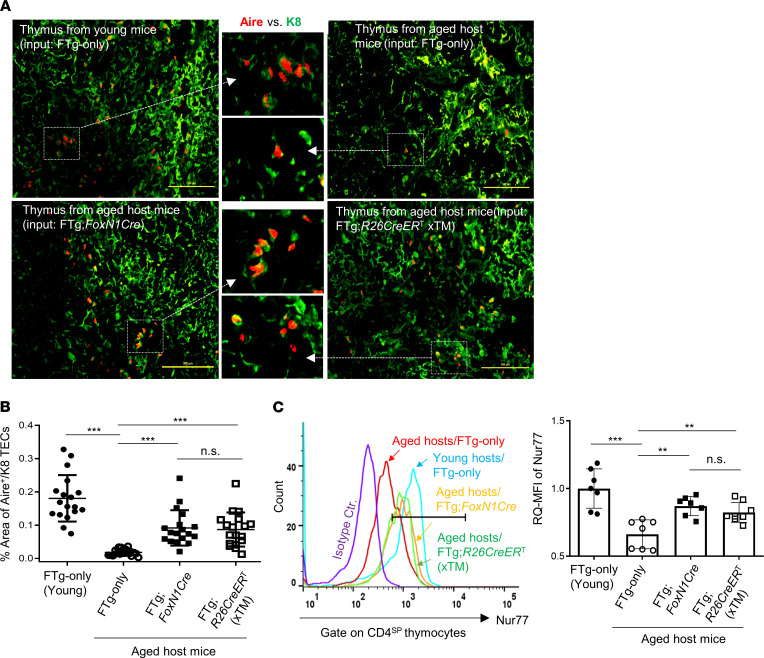
Figure 4. Transplantation of FREFs boosted Aire gene expression in the aged thymus and showed enhanced negative selection signaling strength via Nur77 in CD4SP thymocytes of aged mice.
Same experimental setting as described in Figure 2. (A) Representative immunofluorescence staining images of Aire+ TECs (red) in K8+ TEC counterstaining (green). Data are representative of 3 biological replicates in each group with essentially identical results. Scale bar: 100 μm. (B) Summarized result shows the percentage area of Aire+ TECs against K8+ counterstaining based on the slides in A. Each symbol represents 1 thymic tissue section; 5–7 of these thymic tissue sections per thymus at different physical locations (nonsequential slides) were counted using 3 thymuses per group and analyzed with ImageJ software (NIH; a total of 17–19 thymic tissue sections from 3 individual thymuses per group were observed). (C) Flow cytometric results show increased Nur77 signaling strength (relative quantitative [RQ] mean fluorescence intensity [MFI]) in CD4SP thymocytes of young (control) or aged mice that were engrafted with MEFs or 2 types of FREFs. Left panel: histogram of Nur77 MFI in CD4SP thymocytes; right panel: Nur77 RQ-MFI in CD4SP populations of various groups (n = 7–8 mice per group). All results represent the mean ± SD. The statistical analysis was performed by 1-way Newman-Keuls multiple-comparisons test. **P < 0.01, ***P < 0.001. n.s., not significant.

