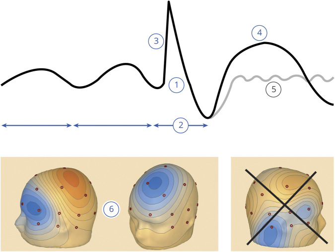
Figure 1. Infographic summarizing the 6 IFCN criteria for identifying IEDs.
(1) Di- or tri-phasic wave with pointed peak; (2) different wave duration than the ongoing background activity; (3) asymmetry of the waveform; (4) followed by a slow after-wave; (5) the background activity is disrupted by the presence of the IEDs; and (6) voltage map with distribution of the negative and positive potentials suggesting a source in the brain corresponding to a radial, oblique, or tangential orientation of the source.23 For further details, see the Methods section. The voltage maps in window (6) show a tangential orientation (source in the left middle frontal gyrus) and a radial orientation (source in the left superior frontal gyrus); the irregular distribution of the potentials in the last voltage map does not imply a source in the brain (it was an artifact with sharp morphology) and does not fulfill this criterion; negative potentials are in blue, and positive potentials are in red. IED = interictal epileptiform discharge; IFCN = International Federation of Clinical Neurophysiology.

