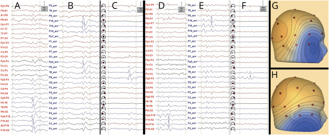
Figure 2. Examples of IED (A–C) and nonepileptiform sharp transient (D–F).
Montages: longitudinal bipolar (A and D), common average (B and E), and source space (C and F). Voltage maps are shown in G and H. The transient shown in A–C with voltage map in G (oblique distribution corresponding to a source in the right temporal pole) fulfills all IFCN criteria in sensor space, except for criterion 5, and hence, it qualifies as an IED; in source space, it also qualifies as an IED, and the propagation from the temporal pole to the basal temporal region can be observed (C). The transient shown in D–F with voltage map in H only fulfills 2 IFCN criteria in sensor space (1 and 6), and hence, it does not qualify as an IED; in source space, one can observe that the transient belongs to the background activity from the right basal temporal region (fragmented during drowsiness); the orientation of the voltage map corresponds to a source in the right basal temporal region (fulfilling criterion 6). Voltage maps are useful in distinguishing IEDs from artifacts (figure 1); however, nonepileptiform transients originating from the brain can show voltage distributions similar to IEDs. IED = interictal epileptiform discharge; IFCN = International Federation of Clinical Neurophysiology.

