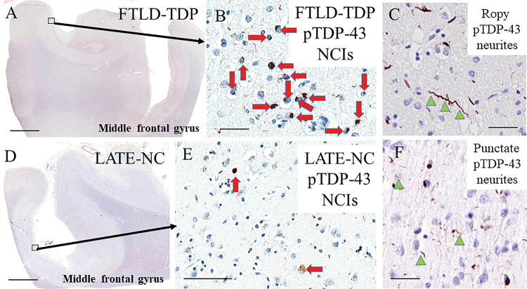
Figure 2.
Representative photomicrographs showing TDP-43 immunoreactive features in LATE-NC and FTLD-TDP. TDP-43 neuropathological features in FTLD-TDP type A/B (A–C) and LATE-NC (D–F) are similar in morphology but frequently differs by severity. Dense neuronal cytoplasmic inclusions (NCIs, red arrows; B) and ropy dystrophic neurites (dystrophic neurites shown with green arrowheads; C) are seen the superficial neocortical layers in FTLD-TDP type A middle frontal gyrus. LATE-NC middle frontal gyrus has milder pathology (E) and punctate dystrophic neurites (F) can be seen in both FTLD-TDP and LATE-NC. Scale bars = 3 mm in A and D; 50 µm in B, C, and F; 100 µm in E.

