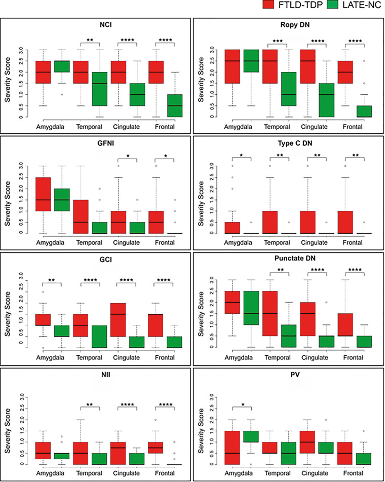
Figure 3.
Scores of TDP-43 proteinopathic features by brain region, stratified by diagnosis of LATE-NC or FTLD. Cortical pathology is more severe in FTLD-TDP (red) compared to LATE-NC cases (green). TDP-43 pathology was scored separately for individual morphologies for each case and region. The burden of TDP-43 pathology in amygdala was similar between severe LATE-NC and FTLD-TDP and was typically characterized by the presence of neuronal cytoplasmic inclusions (NCI), ropy dystrophic neurites (DN) and punctate dystrophic neurites. Cortical pathology in anterior cingulate, superior and middle temporal, and middle frontal gyrus was more limited in LATE-NC. In FTLD-TDP cases, there was typically moderate to severe neuronal cytoplasmic inclusions and ropy dystrophic neurites, which were usually mild to rare to LATE-NC, although these cases were selected to represent the more severe portion of the LATE-NC pathological spectrum. Box and whisker plots show the median (solid line) and whiskers indicate variability outside the upper and lower quartiles (average of datasets from two neuropathologists, UK-1 and UK-2). *P < 0.05, **P < 0.01, ***P < 0.001, ****P < 0.0001 Mann-Whitney-Wilcoxon analysis. DN = various types of TDP-43 immunoreactive dystrophic neurites, including DN characteristic of FTLD type C (Type C DN), ropy DN and punctate DN; GFNI = granulofilamentous neuronal inclusions; NII = neuronal intranuclear inclusion; PV = perivascular TDP-43 proteinopathy; WM GCI = white matter glial cytoplasmic inclusion.

