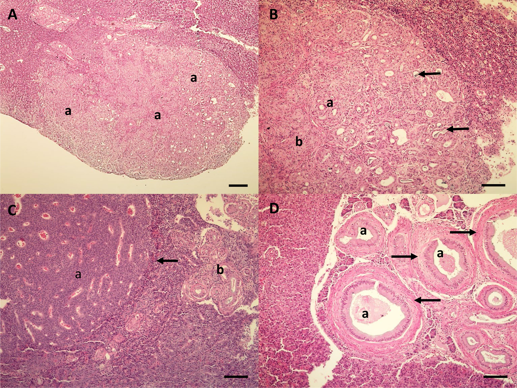
Figure 4.

Microscopic lesions in the liver of white sucker from the St. Louis River Area of Concern. A. Cholangiocarcinoma (a) on the surface of the liver. Scale bar equals 200 μm. B. Higher magnification of cholangiocarcinoma illustrating proliferating bile ducts of varying shape and size. Some areas are well differentiated (a), while other areas (b) have increased atypia and pleomorphism. Some bile ducts have bile present in the lumen (arrows). Scale bar equals 100 μm. C. Hepatic cell adenoma (a) with compression along the periphery of the adenoma (arrow). Bile duct proliferation and fibrosis (b) is also present. Scale bar equals 100 μm. D. Proliferation of bile ducts (a) with fibrosis (arrows). Scale bar equals 100 μm. Hematoxylin and eosin stain.
