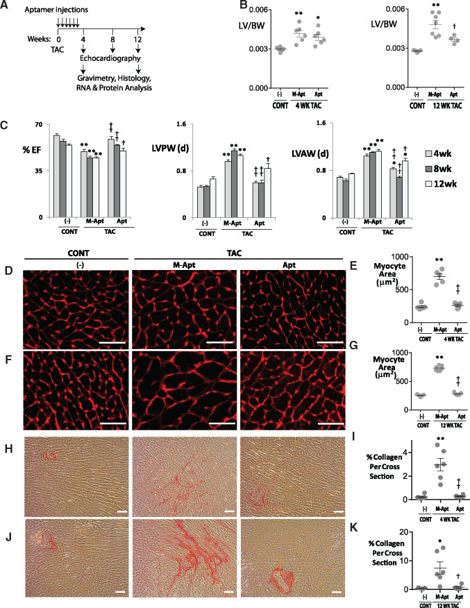
Figure 1.
OPN aptamer prevents cardiac dysfunction, hypertrophy, and fibrosis in TAC mice. (A) Protocol for OPN aptamer therapy in the mouse TAC heart failure model. A total of 10 injections were administered, one subclavian and nine tail vein injections. (B) Effect of OPN aptamer on LV to body weight ratios, and (C) echocardiographic indices. Representative images of Wheat Germ Agglutinin staining and corresponding myocyte area of LV cross sections at 4 (D,E) and 12 weeks (F,G) post-TAC after systemic injections of OPN aptamer (Apt) or mutant OPN aptamer (M-Apt). Representative images of Picrosirius Red staining and corresponding % collagen quantification of LV cross sections at 4 (H,I) and 12 weeks (J,K) post-TAC after systemic injections of OPN aptamer or mutant OPN aptamer. Detailed echocardiographic data and statistics are shown in Supplementary material online, Table SI. Error bars represent SEM. P-values are from ANOVA with Tukey multiple correction. *P ≤ 0.05 vs. Sham; **P ≤ 0.01 vs. Sham; †P ≤ 0.05 vs. TAC + M-Apt; ††P ≤ 0.01 vs. TAC + M-Apt. n , 5–13 mice per group. Cont, sham or no-surgery controls; TAC, Transverse Aortic Constriction; EF, Ejection Fraction; LVPW, LV posterior wall; LVAW, LV anterior wall; Scale bar, 50 μm.

