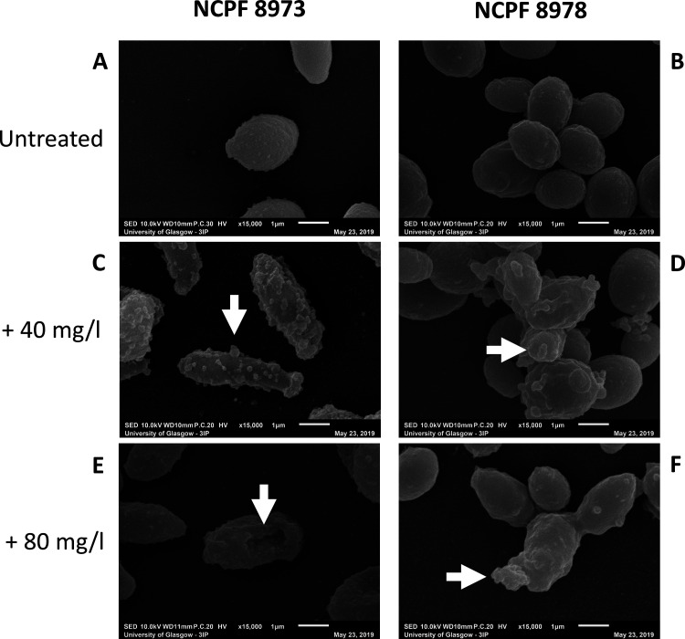
FIG 1.
Scanning electron microscopic images of chitosan-treated Candida auris. Chitosan-treated 24-h biofilms of nonaggregative (non-Agg) NCPF 8973 and aggregative (Agg) NCPF 8978 C. auris were visualized using scanning electron microscopy (SEM). (A and B) Untreated non-Agg and Agg biofilms were used as controls and treated in the same way minus chitosan. Non-Agg and Agg biofilms of C. auris were treated with 40 mg/liter (C and D) or 80 mg/liter (E and F) for 24 h prior to imaging at ×15,000 magnification. White arrows highlight the encapsulation of C. auris cells by chitosan particles and deflation in cell morphology of the non-Agg NCPF 8973 isolate (E).

