
FIGURE 4.
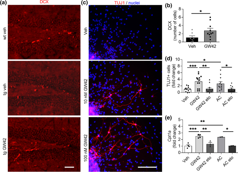
GW0742 treatment increases neurogenesis in vivo and in vitro in conjunction with elevated Cpt1a. (a) Representative immunohistochemistry images of hippocampal slices from wild type (wt) and transgenic (tg) APP/PS1-mice treated with vehicle or 30 mg/kg GW0742 for 14 days and stained for newborn neurons with DCX (red). Scale bars, 100 μm. (b) Corresponding graph for the number of DCX positive cells per hippocampal slice counted from six brain sections at400-μm intervals. n = 7–10. (c) Representative immunocytochemistry images of wt neurospheres differentiated for73 days in vitro and treated with vehicle, 10 or 100 nM GW0742 and stained for mature neurons with TUJ1 (red) and for nuclei (blue). Scale bars, 100 μm. (d) Fold change in the number of TUJ1-positive cells after neurosphere differentiation and treatment for 3 days with 100 nM GW0742, 200 μM etomoxir (eto) to block the activity of CPT1, or 300 μM acylcarnitine (AC) to provide a direct substrate for fatty acid oxidation (FAO). n = 10–14. (e) Corresponding relative gene expression levels of Cpt1a shown as a fold change to vehicle. n = 2–4 biological replicates in independent experiments. All data are presented as mean ± SEM with *p < .05, **p < .01, ***p < .001 as analyzed by t test or two-way anova followed by Bonferroni post hoc test