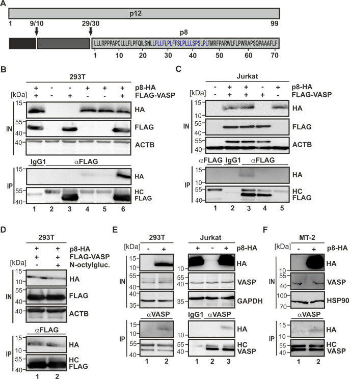
Fig 1. VASP is a novel interaction partner of p8.
(A) Scheme of the HTLV-1 p8 protein and its precursor p12. A motif mediating potential interactions with the VASP EVH1 domain is highlighted in blue. (B) 293T cells or (C) Jurkat T-cells were transfected with expression plasmids p8-HA and FLAG-VASP or empty control vectors. After 48 h, cells were lysed and 10% of the lysates were taken as input (IN). Co-immunoprecipitations (IPs) were performed using anti-FLAG antibodies or isotype-matched control antibodies (IgG). Immunoblots are shown. HC, heavy chain. (D) 293T cells were transfected with expression plasmids p8-HA and FLAG-VASP. After 48 h, cells were lysed and 10% of the lysates were taken as input. Lysates were treated without (lane 1) or with 60 mM N-octyl-β-D-glucoside (N-ocytlgluc.; lane 2). IPs were performed using anti-FLAG antibodies. Immunoblots are shown. (E) 293T, Jurkat or (F) HTLV-1-infected MT-2 cells were transfected with expression plasmids p8-HA. After 48 h, cells were lysed and 10% of the lysates were taken as input. Endogenous VASP was precipitated using anti-VASP antibodies. Immunoblots are shown.

