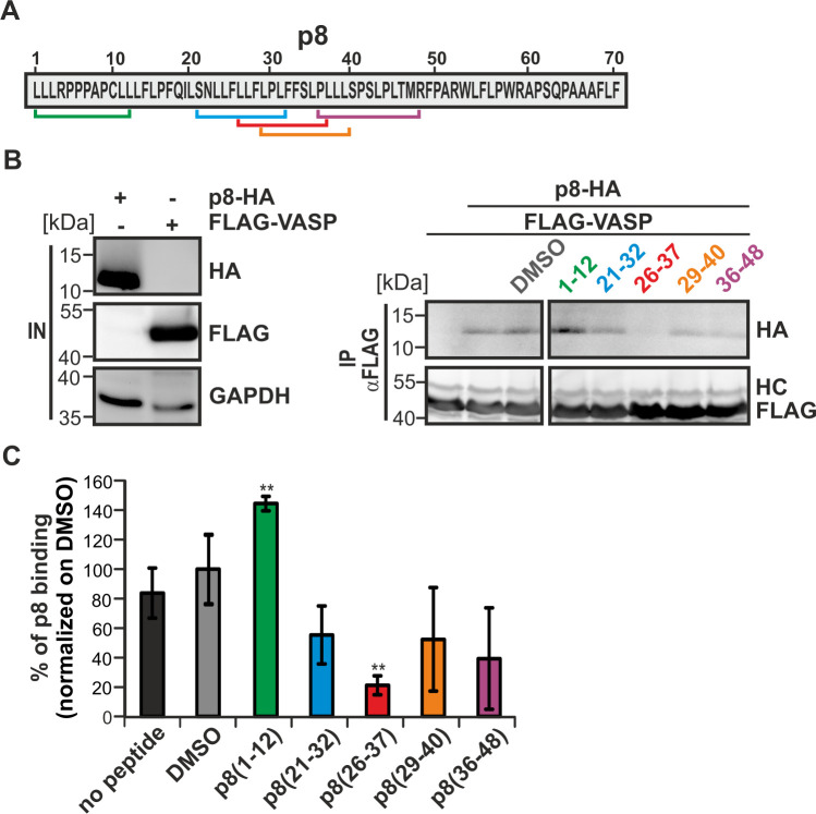
Fig 3. p8-mimicking peptides reduce the interaction between p8 and VASP.
(A) Amino acid sequence of p8. Bars indicate location of competitive p8 inhibitory peptides. (B) p8-HA and FLAG-VASP were expressed individually in 293T cells. After 48 h, cells were lysed and 10% of the cell lysate were taken as input (IN). The remaining lysates were co-incubated with the peptides shown in (A), or with the solvent control DMSO for 1.5 h at 20°C. Thereafter, lysates were mixed for 1.5 h, and co-precipitations (anti-FLAG) were performed (IP). Representative immunoblots are shown. The blot (IP) was cut due to technical reasons. HC, heavy chain. (C) The impact of p8-mimicking peptides on binding of p8-HA after precipitation of FLAG-VASP was assessed by densitometry. DMSO was set as 100%. The means of four independent experiments ± SE were compared to DMSO-treated cells using Student’s t-test. ** indicates p<0.01.

