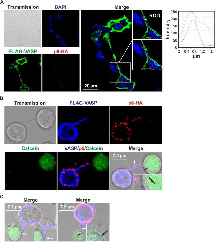
Fig 5. p8 and VASP partially co-localize in protrusive structures between cells and p8 is transferred to target T-cells via VASP-containing protrusions.
(A) Confocal laser scanning microscopy of 293T cells upon co-transfection of p8-HA and FLAG-VASP expression plasmids. Stains of FLAG-VASP (green), p8-HA (red), the cell nuclei (DAPI, blue), the merge of all three stains and transmitted light are shown. Regions of interest (ROI) within a protrusive structure are shown and graphs show the fluorescence intensities of FLAG-VASP- and p8-HA-specific fluorescence along the ROI. (B) Jurkat T-cells were co-transfected with expression plasmids p8-HA, FLAG-VASP and pMACS-LNGFR. After 48 h, transfected cells were enriched by magnetic separation using LNGFR-specific microbeads and co-cultured with untransfected Jurkat T-cells pre-stained with the live cell marker Calcein (green) on poly-L-lysine-coated coverslips for 30 min at 37°C. Immunofluorescence stainings of FLAG-VASP (blue), p8-HA (red), the merge of all stainings and transmitted light are depicted. White arrow: p8 co-localizing with VASP in a protrusion; black arrow: p8 in co-cultured target Jurkat T-cell. (C) Two additional examples of merges showing p8 co-localizing with VASP in a protrusion (white arrows) and p8 in co-cultured target Jurkat T-cells (black arrows) as described in (B).

