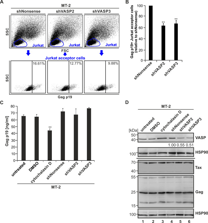
Fig 10. Repression of VASP impairs virus transmission to uninfected T-cells, but not Gag processing and virus release in chronically infected MT-2 cells.
(A) Jurkat T-cells were co-cultured with stably transduced MT-2 cells (shNonsense, shVASP2, shVASP3) at a ratio of 1:1 for 1 h. The transfer of Gag p19 to Jurkat acceptor cells was measured by flow cytometry using the primary antibodies mouse anti-Gag p19 and the secondary antibodies anti-mouse Alexa Fluor 647. Upper part: Jurkat acceptor cells were gated by forward scatter (FSC) and side scatter (SSC) analysis; lower part: Gag p19-specific fluorescence (indicated by numbers) in Jurkat acceptor T-cells. (B) The amount of Gag p19 positive Jurkat acceptor cells normalized on the MT-2/shNonsense control cells of four independent experiments ± SE is depicted and was compared using Student’s t-test (**, p<0.01). (C) The amount of Gag p19 protein in the supernatant of transduced MT-2 cell lines was assessed by Gag p19 ELISA. MT-2 cells treated with 5 μM cytochalasin D, an inhibitor of actin-polymerization, in comparison to the DMSO solvent control served as positive control for an impaired Gag p19 release. The mean of four independent experiments ± SE is depicted and was compared using Student’s t-test (**, p<0.01). (D) Western blot analysis depicting VASP, Tax and Gag p55 precursor and processed Gag p19 matrix protein in stably transduced MT-2 cells (shNonsense, shVASP2, shVASP3) and the indicated controls. Numbers indicate densitometric analysis of VASP normalized on Hsp90 and shNonsense.

