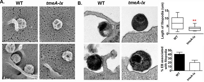
Fig 6. TmeA is partially required for chlamydial association with filopodia.
Human cervical cells were infected for 15min at a MOI of 50 with wild-type or tmeA-lx and imaged by scanning (A) or transmission (B) electron microscopy. Quantification of EBs associated with filopodia was assessed from two independent experiments with 50 EBs counted per experiment. Length of surface structures were measured to confirm filopodia (top). Bacteria associated with filopodia were compared to total bacteria to determine percent associated with filopodia. Two representative images for each are shown. Scale bar is 200nm.

