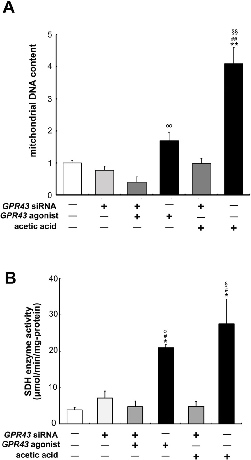
Fig 8. Proliferation of mitochondria by the treatment of acetic acid or GPR43 agonist through the activation of GPR43.

(A) Genomic DNA was extracted from L6 cells that were transfected or non-transfected with GPR43- siRNA after the treatment or no treatment of 0.5 mM acetic acid or 1.0 μM GPR43 agonist for 30 min. Real-time PCR analysis was carried out for the determination of mt-Nd1 level in L6 myotube cells. (B) SDH activity (nmol/min/mg of protein) in L6 cells of same condition with (A) was measured by consumption rate of DCPIP as described in Materials and Methods. Each bar represents the mean ± SE (n = 3–6). Multiple comparisons were analyzed with one-way ANOVA followed by the Tukey-Kramer post hoc test. Statistical differences are shown as *p< 0.05, **p< 0.01, compared with non-treated control; #p< 0.05, ##p< 0.01, compared with GPR43 siRNA; §p< 0.05, §§p< 0.01, compared with GPR43 siRNA + ace; op< 0.05, oop< 0.01, compared with GPR43 siRNA + agonist.
