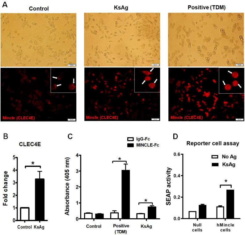
Fig. 4.
KsAg-Mincle-Fc fusion protein binding and subsequent Mincle-dependent SEAP release. (A) Immunofluorescence image of Mincle protein. White arrows indicate Mincle expression on the cells. (B) CLEC4E mRNA expression (*P < 0.05). (C) KsAg-Mincle-Fc fusion protein binding. Trehalose-6, 6’-dimycolate (TDM, a well-known Mincle ligand) was used as a positive control (*P < 0.05). (D) Difference of NF-κB-induced SEAP activity between HEK-BlueTM hMincle and HEK-BlueTM Null1-v cells (parental cell line) with and without KsAg treatment. Data are expressed as the mean ± SD. There was a significant difference between HEK-BlueTM hMincle cells that were treated with KsAg and those that were untreated (*P < 0.001).

